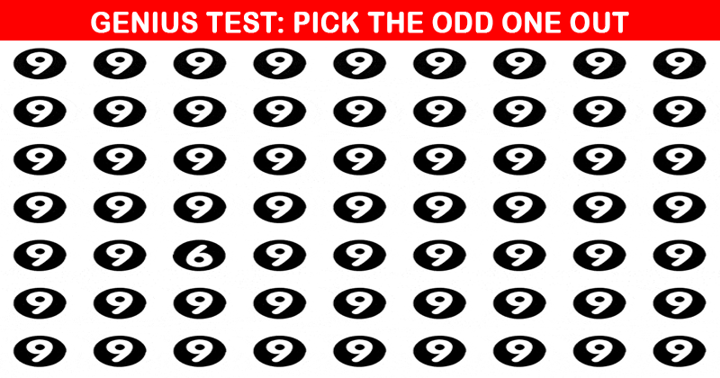
You have to ace both levels to pass this eye test. Give it a try now!
Your medical history
Your doctor will ask you about your vision and your general health. They will ask about:
- your family’s medical history,
- what medications you take, and
- whether you wear corrective lenses.
Your visual acuity
This is the part of an eye exam people are most familiar with. You will read an eye chart to determine how well you see at various distances. You cover one eye while the other is being tested. This exam will determine whether you have 20/20 vision or not.
Your prescription for corrective lenses
Your doctor will ask you to look at an eye chart through a device called a phoroptor. The phoroptor contains different lenses. It will help determine the best eyeglass or contact lens prescription for you.
Your pupils
Your doctor may check how your pupils respond to light by shining a bright beam of light into your eye. Pupils usually respond by getting smaller. If your pupils widen or don’t respond, this may reveal an underlying problem.
Your side vision
Loss of side vision (peripheral vision) may be a symptom of glaucoma. This test can find eye problems you aren’t aware of because you can lose side vision without noticing.
Your eye movement
A test called ocular motility evaluates the movement of your eyes. Your ophthalmologist looks to see if your eyes are aligned. They also check that your eye muscles are working properly.
Your eye pressure
Eye pressure testing, called tonometry, measures the pressure within your eye (intraocular eye pressure, or IOP). Elevated IOP is one sign of glaucoma. The test may involve a quick puff of air onto the eye or gently applying a pressure-sensitive tip near or against your eye. Your ophthalmologist may use numbing eye drops for this test for your comfort.
The front part of your eye
Your ophthalmologist uses a slit-lamp microscope to light up the front part of the eye. This includes the eyelids, cornea, iris and lens. This test checks for cataracts or any scars or scratches on your cornea.
Your retina and optic nerve
Your ophthalmologist will put dilating eye drops in your eye to dilate, or widen, your pupil. This will allow them to examine your retina and optic nerve for signs of damage from disease. Your eyes might be sensitive to light for a few hours after dilation.